
Description:
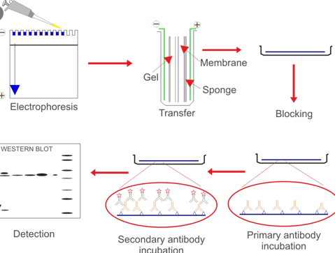
A western blot is a way used to determine the presence of an antigen in a selected tissue homogenate or protein extract. Typically, protein samples are resolved by their dimension by gel electrophoresis and transferred onto a membrane. The membrane is then probed with an antigen particular antibody which is then itself detected utilizing an enzyme conjugated antibody. Enzyme substrate is then utilized and the membrane is visualized for the presence of sign. see instance western blots from our validation program!
Procedure:
Please seek the advice of the product information sheet for the suitable focus of main antibody and another particular situations.
Cell Lysate Preparation:
A. Reagents and Materials
- Phosphate Buffered Saline; 1X PBS, pH 7.4
- SDS-Loading Buffer: 0.05M Tris-HCl, pH6.8, 0.1M DTT, 2percentSDS, 10% glycerol, 0.1% bromophenol blue
- Cell Detaching Trypsin Buffer: 0.25% trypsin in 0.53M EDTA answer
- Cell Lysis Buffer (1% NP40, 0.5% DOC, 1mM EDTA , 65mM Tris-HCl pH=6.4, 1mM PMSF, 1ug/ml Aprotinin, 1ug/ml Leupeptin, 1ug/ml Pepstatin)
- BCA Protein Assay Kit
B. Equipment
- Ultrasonicator
C. Protocol
- Cell tradition and harvest
a) Cell subculture
b) Incubate cells for about three days and rely the cells. When the focus attain 106 cells/ml, detach cells, spin down and wash twice with 1XPBS .
c) Add cell lysis buffer to cell extracts (1ml lysis buffer /5×107 cells). Vortex the combination for 5min and retailer at -20C. - Preparation of tissue extracts
a) Thaw the frozen, uncooked tissue pattern by vortexing and repeat the freeze-thaw cycle twice. - b) Disrupt tissue with ultrasonicator. Keep all the operation on ice.
- Measure focus of cell samples
a) Transfer contents to a microcentrifuge tube. Centrifuge at 12,000 rpm for 15 minutes. Then accumulate supernatant into an appropriately labeled tube (for the detection of membrane sure proteins, use the insoluble, cell pellet). Dilute the supernatant lysate and the entire lysate with out centrifugation at 1:4, 1:Eight and 1:16 with 1XPBS. Measure protein focus utilizing cell lysis-compatible protein assay (BCA protein assay). - b) Mix the cell lysate with SDS loading buffer to make the specified last focus. Incubate the lysates at 100°C for 10min.
- Quality management
a) Test cell lysate by SDS-PAGE. The cell lysate is evaluated as certified, if the bands are clear and haven’t any apparent smear. - b) Test cell lysate by Western Blot. The main antibody we used is the antibody towards the marker proteins. The cell lysate is evaluated as certified, if the WB picture reveals 5 bands.
SDS-PAGE
A. Reagents and Materials
- Stacking gel buffer: 0.5M Tris-HCl, pH6.8, 0.4% SDS
- Separating gel buffer: 1.5M Tris-HCl, pH8.8, 0.4% SDS
- Acrylamide inventory answer: 29% acrylamide plus 1.0% bis-acrylamide
- 10% ammonium persulfate
- TEMED(N,N,N’,N’-tetramethylene-ethylenediamine)
- Electrophoresis buffer: 0.25M Tris Base, 2M Glycine, 1% SDS
- Coomassie gel stain answer: 0.1% Coomassie blue R-250, 30% ethanol, 10% acetic acid.
- Coomassie gel destain answer: 30% ethanol, 10% acetic acid.
B. Equiment
- Minigel equipment: Bio-Rad Mini-Protean 3 Dodeca Cell
- Power provide: Biio-Rad PowerPac HC
- Gradient gel former: Bio-Rad Model 485 Gradient Former
C. Protocol
- Assemble mutil-casting chamber in accordance with the producer’s directions. All following operations associated with equipment ought to observe manufacture’s directions.
- Make separating gel as following system in an acceptable beaker. Ammonium persulfate and TEMED ought to be added earlier than pouring gel.
| Separating focus | 8% | 10% | 12% | 15% |
|---|---|---|---|---|
| Separating gel buffer | 16 | 16 | 16 | 16 |
| Acrylamide inventory answer | 16 | 20 | 24 | 30 |
| H2O | 27.4 | 23.4 | 19.4 | 13.4 |
| 10% ammonium persulfate | 0.6 | 0.6 | 0.6 | 0.6 |
| TEMED | 0.05 | 0.05 | 0.05 | 0.05 |
b) For making 12 gradient gels (ml):
| Separating focus | Low | High | ||
| 6% | 10% | 18% | 20% | |
| Separating gel buffer | 8 | 8 | 8 | 8 |
| Acrylamide inventory answer | 6 | 10 | 18 | 20 |
| H2O | 15.5 | 11.7 | 3.7 | 1.6 |
| 10% ammonium persulfate | 0.3 | 0.3 | 0.3 | 0.3 |
| TEMED | 0.025 | 0.025 | 0.025 | 0.025 |
Use gradient gel former to make gradient gel. Use 10-20% gel for low molecular weight (MW< 20kD) protein identification and use 6-18% gel for prime molecular weight (MW>100kD) protein identification.
- Pour the separating gel
- Pour the stacking gel
- Loading samples
- Running gel
- Processing Gel for various makes use of
- a) Staining gel with Coomassie Blue. If separated proteins are wanted to be seen within the gel instantly, stain gel with Coomassie gel stain answer with agitation for 40 minutes after which destain with Coomassie gel destain answer till background staining disappears.
- b) Immunoblotting: If particular protein is required to be detected by antibody, course of gel as described within the protocol of Gel Transfer and Western Blot.
Gel Transfer
A. Reagents and Materials
- Transfer buffer 10X inventory answer: 0.25M Tris, 2M Glycine. Add methanol to 10% after dilution to 1X buffer and simply earlier than use.
- Blocking buffer: 5.0% non-fat dry milk in 1 X PBS, pH=7.4.
- PVDF membrane
B. Equipment
- Electroblotting equipment: Bio-Rad Trans-Blot Cell
- Power provide: Bio -Rad PowerPac H
C. Protocol
This protocol is the next steps of SDS-PAGE.
- PVDF membrane course of: Cut PVDF membrane to the identical dimension of SDS-PAGE gel. Soak membrane sequentially in 100% methanol for 15 seconds, distilled water for five seconds and into switch buffer for no less than 10 minutes.
- Electrotransfer
- a) Arrange gel-membrane sandwich as described in producer’s instruction. Place the switch sandwich unit into buffer tank, fill with pre-cooled switch buffer and fix the electrodes. Set the facility provide to 100V and switch for 80 minutes.
- Blocking
- a) Disconnect switch equipment, take away switch cassette and switch PVDF membrane to blocking buffer. Rock the blocking membrane on shaker for two hours at RT or preserve it at 4C in a single day.
- b) Follow Immunoblotting protocol as described.
Immunoblotting
A. Reagents and Materials
- Antibody dilution buffer: 5% non-fat dry milk in 0.1% PBST
- Developing answer/Fixative answer for X-ray movie
- Membrane washing buffer: 0.1% PBST
- Primary antibody; Aviva affords >30,000 antibodies for Western Blot
- Secondary antibody; Goat anti-Rabbit I gG-HRP (CAT#ASP00001)
- Horseradish Peroxidase Substrate: e.g. Chemiluminescent FemtoMax (CAT# OORA01695)
- X-ray movie
B. Equipment
- Rotary shaker
C. Protocol
This protocol is to be adopted after Gel Transfer.
- Primary antibodies preparation
a) The last main antibody focus ought to be 1.25 – 5.Zero ug/ml for protein A purified IgG (_T100) and 0.25 – 1.Zero ug/ml for peptide purified IgG (_P050). See product particulars for actual dilution to make use of. - Pretreat membrane: Wash the membrane in wash buffer for 10min.
- Primary antibodies incubation
- a) Dilute the first antibody in antibody dilution buffer to acceptable antibody focus, then incubate for 1hour at room temperature with agitation to allow enough homogeneous protecting of the membrane and stop uneven binding. Overnight at 4C with agitation can be acceptable.
- Wash membrane: Pour off main antibody answer and wash membrane 6 occasions for five minutes every time in Membrane washing buffer.
- Secondary antibodies incubation
- Wash membrane: Pour off main antibody answer and wash membrane 6 occasions and for five minutes every time in Membrane washing buffer.
- Development
- Exposure in darkroom
- a) Exposure the membranes to X-ray movie in cassette for 1 minute. Manual movie growth is used to regulate the incubation time of the x-ray movie within the creating answer and fixative answer. Normally, about 2-10 min for growth and 5 min for fixation. Dry the movie after totally washing with water.